|
|
|
|
|
|
|
|
|
|
|
|
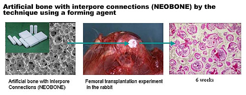
Ⅰ Bone regeneration using artificial bone hybridized with bone marrow
stem cells |
|
|
|
For bone tissue regeneration, biomaterial serving as a good scaffold
and osteoblasts with osteogenicity are necessary. In this study, novel
hydroxyapatite with a continuous pore structure with properties physically
and biologically equivalent to those of bone will be developed by applying
the technique of forming ceramics with completely continuous pores using
a foaming agent. This novel hydroxyapatite will be hybridized with osteogenic
cells, such as auto-bone marrow cells and bone morphogenetic proteins or
their genes to develop artificial bone with biological activity capable
of complementing bone defect with a specific size in a specific region.
Artificial bone with a shape corresponding to bone defects will be prepared by morphological simulation technique using a computer. For a large bone defect, the hydroxyapatite will be implanted with vascular tissues in muscle tissue in vivo to prepare artificial bone with nutrient blood vessels transplantable for a specific region. |
|
|
 |
|
|
|
|
|
 |
|
|
|
|
|
|
|
|
|
|
|
|
|
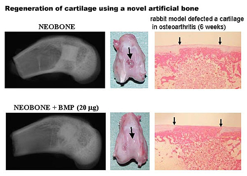
Ⅱ Regeneration of articular cartilage using synovial membrane-derived mesenchymal cells |
|
|
|
For regeneration and repair of articular cartilage, auto-chondrocytes
and bone marrow stem cells have been used, but good therapeutic results
have not been obtained. In this study, we will develop batch culture technique
of synovial cells and technique of preparing 3-dimentional synovial cell-matrix
complex with size and shape corresponding to therapeutic uses.
Since the complex matrix can be constructed into various shapes and has sufficient physical strength, its surgical manipulation is easy. Since matrices contain abundant cell adhesion factors such as collagen and fibronectin, the complex and the recipient tissue are biologically connected within a short time.
Using this 3-dimensional synovial cell-matrix complex, we will attempt
clinical regeneration of articular cartilage for cartilage injury and degenerated
cartilage in osteoarthritis after animal studies in rabbits and pigs at
the Medical Center for Translational Research of the Hospital of Osaka
University School of Medicine. |
|
|
|
|
|
|
|
|
|
|
|
|
|
|
|
|
|
|
|
|
|
|
Ⅲ Development of novel joint prosthesis with metal surface processed
by YAG laser scanning |
|
|
|
The loosening of a human joint prosthesis from a bone is one of the
most serious problems which have not been cleared. Conventional porous
coating method on a metal surface, which is widely available in the human
joint prosthesis, cannot provide a microscopically idealistic shape of
the metal surface for the bonding between the surface and a bone. The method
is used to make rough, but it cannot precisely regulate the shape. Our
method using a YAG laser scanning is originally developed to treat a metal
surface in Osaka Univ. Graduate School. It can provide any microstructure
of the metal surface.
The microscopic canal structures treated on the metal surface had more
than twice of the bonding strength compared to a porous coated surface.
And a new bone ingrowth was histologically observed in all cavities of
the canals. The results encourage the development of metal surface treatment
to make a new joint prosthesis which has no loosening from a bone. |
|
|
 |
|
|
|
|
|
|
Other study contents of Yoshikawa laboratory |
|
|
|
|
■Rheumatoid arthritis therapy for using NFkB decoy nucleic acid pharmaceuticals
■Development of a new diagnostic method of bone soft tissue tumors using
gene analysis
■Analysis of the kinematics after joint replacement using 3-dimensional
CT and MRI
■Development of a robotic surgical method of artificial joint
|
|
|
|
|
Yoshikawa associated page≫ |
|
|
|
|
|
|
|